Whole-Body Pharmacokinetics of Lipid, mRNA and Translated Protein Following Intravenous Administration of Spike Protein Expressing mRNA-LNP in Mice
- Apr 25
- 8 min read
Technical Review | Pharmaceutical Research, 2026 | Kumar, Tiwari, Rajwade, Kulkarni, Patel, Shah et al. | Featured Tool: PreciGenome NanoGenerator Flex-M®
Background
Lipid nanoparticle (LNP)-encapsulated mRNAs have emerged as a transformative platform for prophylactic and therapeutic applications, as demonstrated by the clinical success of COVID-19 vaccines such as Comirnaty (Pfizer-BioNTech) and Spikevax (Moderna). While extensive research has focused on optimizing LNP formulations for potency and immunogenicity, a critical gap remains in understanding the pharmacokinetics (PK) of the individual components—the lipid carrier, the mRNA payload, and the expressed protein—across different tissues.
The dose-exposure-response relationship for mRNA-LNP therapies is more complex than that of traditional drugs. The biodistribution and endosomal escape capability of the lipid nanoparticle dictates mRNA delivery efficiency in vivo. Once inside cells, mRNA half-life and translational efficiency are influenced by structural elements such as untranslated regions, the poly-A tail, 5’ cap, and modified nucleotides. The expressed protein then follows its own intrinsic PK behavior, creating a multilayered system where each component exhibits distinct kinetics.
Most prior studies have evaluated mRNA-LNP biodistribution using reporter mRNAs in a qualitative or semi-quantitative manner, often in limited tissues or relying on labeling strategies that can provide misleading PK data. In this study, Kumar et al. from the University at Buffalo addressed this critical knowledge gap by performing a comprehensive, quantitative whole-body PK study of all three mRNA-LNP components—the ionizable lipid ALC-0315, the encapsulated spike protein mRNA, and the translated spike protein—in plasma and ten different tissues following intravenous administration in mice.
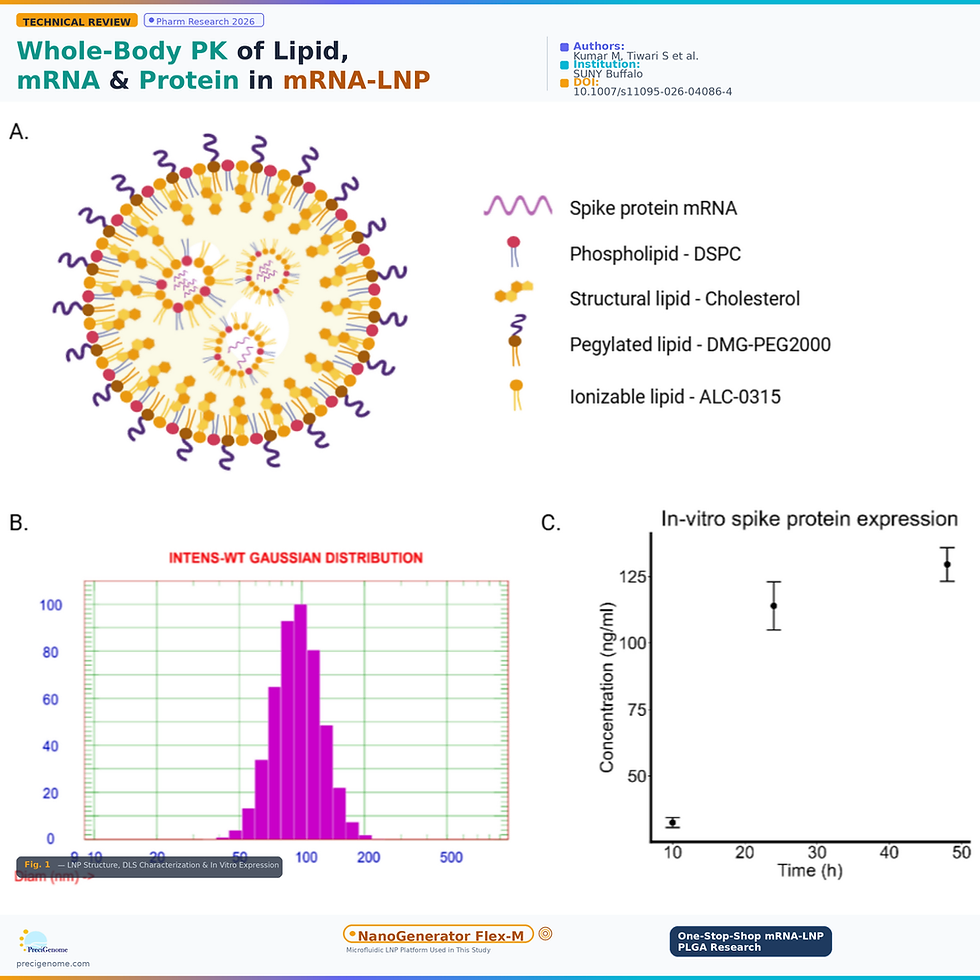
Figure 1. (A) Schematic illustration of the LNP structure. ALC-0315 becomes positively charged at acidic pH during formation, complexing with negatively charged spike protein mRNA in the core. Phospholipids, cholesterol, and PEG-lipids form a stable outer shell. (B) DLS analysis of produced mRNA-LNP revealed a mean diameter of 98 nm and a homogenous formulation with PDI of 0.07. (C) In vitro protein expression kinetics in HEK293T cells following transfection with 500 ng mRNA-LNP, confirming efficient expression of full-length, properly folded spike protein. (Adapted from Kumar et al., Pharmaceutical Research, 2026)
Methodology
Spike protein-encoding mRNA was synthesized via in vitro transcription using the HiScribe T7 mRNA Kit, incorporating CleanCap Reagent AG and N1-methylpseudouridine (m1Ψ) for capping, tailing, and chemical modification. The mRNA was encapsulated within LNPs by microfluidic mixing using the PreciGenome NanoGenerator Flex-M microfluidic mixer. The lipid mixture consisted of ALC-0315, DSPC, cholesterol, and DMG-PEG-2000 at a molar ratio of 50:10:38.5:1.5, dissolved in ethanol. The mRNA was dissolved in 50 mM citrate buffer (pH 4). Lipid and mRNA solutions were rapidly mixed at a volumetric flow rate ratio of 3:1 (aqueous:organic) and a total flow rate of 5 mL/min, with an N:P ratio of 6:1. The resulting LNPs were dialyzed against PBS (pH 7.4), concentrated, and sterile filtered. DLS characterization confirmed a mean diameter of 98 nm with a PDI of 0.07. mRNA-LNP was administered intravenously at 2 mg/kg in C57BL/6 mice. ALC-0315 was quantified by LC-MS/MS, mRNA by RT-qPCR, and expressed spike protein by ELISA across plasma and ten tissues (heart, lung, liver, spleen, kidney, small intestine, large intestine, muscle, brain, and lymph node) over a 14-day time course.

Figure 2. Schematic illustration of the whole-body pharmacokinetic study design. C57BL/6 mice received a single 2 mg/kg IV dose of spike protein mRNA-LNP. Plasma and tissues were collected at multiple time points over 14 days. ALC-0315 lipid was quantified by LC-MS/MS, mRNA by RT-qPCR, and spike protein by ELISA. Anti-spike IgM and IgG titers were also measured. (Adapted from Kumar et al., Pharmaceutical Research, 2026)
Ionizable Lipid ALC-0315 Persists in Tissues for Weeks
Following intravenous administration, ALC-0315 was rapidly cleared from plasma but exhibited prolonged retention in tissues. The lipid reached peak tissue concentrations (Tmax) as early as 10 minutes in most organs, with delayed peaks at 1 hour in liver, spleen, and intestine. The highest tissue exposure, measured by AUC0-t, was observed in the liver (5.76 × 10^7 nM·h), spleen (9.98 × 10^6 nM·h), and kidney (2.55 × 10^6 nM·h), with tissue-to-plasma AUC ratios of 37.09, 6.43, and 1.64, respectively. The lowest exposures were observed in muscle and brain, with nearly 2,000-fold difference in ALC-0315 exposure between liver and brain, highlighting the strong hepatic tropism of ALC-0315-containing LNPs. Substantial ALC-0315 levels were still detectable in tissues two weeks post-dosing, underscoring the prolonged retention of this synthetic ionizable lipid.

Figure 3. Whole-body PK of ALC-0315. (A) Biodistribution of ALC-0315 in major tissues and plasma following IV dosing of 2 mg/kg mRNA-LNP in C57BL/6 mice. Solid circles represent mean concentration; error bars indicate geometric SD. (B) Mean tissue/plasma (T/P) concentration ratios of ALC-0315. Horizontal dashed line represents ratio of 1. Significant accumulation was observed in all tissues, with T/P ratios crossing 1 in all tissues except brain. (Adapted from Kumar et al., Pharmaceutical Research, 2026)
Encapsulated mRNA Shows Rapid Tissue Uptake but Faster Clearance
Unlike ALC-0315, which persisted in tissues for weeks, encapsulated mRNA was cleared much faster, becoming undetectable beyond 72 hours. mRNA was rapidly taken up by tissues within minutes of administration, with Tmax at 10 minutes in most tissues and delayed peaks at 1 hour in liver, spleen, and heart. The highest mRNA exposure (AUC0-72h) was observed in spleen (11.71 nM·h), followed by blood (10.01 nM·h) and liver (7.63 nM·h), with tissue-to-blood AUC ratios of 1.17 and 0.76, respectively. Minimal mRNA distribution was observed in intestine, muscle, and brain (tissue-to-blood AUC ratios <0.05). Notably, while liver had higher mRNA uptake at early time points, its concentration declined more rapidly compared to spleen, resulting in the spleen exhibiting the highest mRNA levels beyond 6 hours—a finding with important implications for spleen-targeted therapeutic applications.

Figure 4. Whole-body PK of mRNA. (A) PK profiles of mRNA in major tissues and plasma following IV dosing of 2 mg/kg mRNA-LNP in C57BL/6 mice. Solid circles represent mean concentration; error bars indicate geometric SD. (B) Mean tissue/blood (T/B) concentration ratios of mRNA. Horizontal dashed line represents T/B ratio of 1. Maximum mRNA localization was observed in spleen and liver. (Adapted from Kumar et al., Pharmaceutical Research, 2026)
Spike Protein Expression Detected Within Minutes of Dosing
Spike protein expression was detected remarkably quickly across all examined tissues, with quantifiable levels within 1 hour post-dosing and liver showing detectable protein as early as 10 minutes—illustrating the speed of LNP uptake, mRNA release, and protein translation. Expression peaked at 6 hours in all tissues, followed by gradual decline. The liver exhibited the highest cumulative expression (AUEC of 574.13 nM·h, tissue-to-plasma ratio of 10), followed by spleen (368.2 nM·h), heart (184.5 nM·h), kidney (168.6 nM·h), and lung (155.1 nM·h). Spike protein expression in large intestine, muscle, and brain was minimal. As spike protein is membrane-bound, low levels in plasma likely resulted from shedding, with plasma showing distinctly slower elimination than tissues—indicating ongoing shedding at later time points.

Figure 5. Whole-body distribution of translated spike protein. (A) Biodistribution of spike protein in major tissues and plasma following IV dosing of 2 mg/kg mRNA-LNP in C57BL/6 mice. Solid circles represent mean concentration; error bars indicate geometric SD. (B) Mean tissue/plasma (T/P) concentration ratios of spike protein. Values greater than 1 at early timepoints indicate production and localization of spike protein within tissues. At later time points, T/P ratios fall below 1 as protein is cleared and shed into plasma. (Adapted from Kumar et al., Pharmaceutical Research, 2026)
Distinct PK Profiles Across All Three Components
A direct comparison of the PK profiles for ALC-0315, mRNA, and spike protein revealed markedly distinct kinetics for each component. ALC-0315 exhibited prolonged tissue retention with the highest accumulation in liver, while mRNA showed the slowest clearance in spleen. Spike protein expression peaked in liver followed by spleen, and was cleared from tissues much faster than either lipid or mRNA. Critically, plasma PK for all three analytes differed significantly from tissue PK, indicating that plasma measurements alone cannot reliably serve as surrogates for tissue exposure. These findings underscore the importance of evaluating all three components independently to fully understand mRNA-LNP pharmacology.

Figure 6. Pharmacokinetic profiles of (A) ALC-0315 lipid, (B) mRNA, and pharmacodynamic profile of (C) expressed spike protein across plasma/blood and major tissues. Profiles are overlaid to compare biodistribution and expression kinetics. ALC-0315 LNPs exhibited predominant accumulation in the liver, indicating strong hepatic tropism. mRNA showed the slowest clearance in the spleen. Spike protein expression peaked in the liver, followed by the spleen. (Adapted from Kumar et al., Pharmaceutical Research, 2026)
Robust Humoral Immune Response Against Spike Protein
Systemic administration of spike protein mRNA-LNP elicited a rapid and robust humoral immune response. Anti-spike IgM titers were first detectable at 24 hours post-dosing at low titers (1:10), increasing markedly by 72 hours (1:1000) and remaining elevated through days 7 and 14. Anti-spike IgG was first detected on day 7 at a titer of 1:1000, indicating initiation of class-switched antibody production. By day 14, IgG titers had increased to 1:10,000, reflecting continued expansion and affinity maturation of antigen-specific B cells. The rapid decline in spike protein concentrations after 72 hours aligned with the onset of anti-spike antibody responses, suggesting a potential role for immune-mediated elimination of the expressed protein.

Figure 7. Anti-spike protein humoral immune response. (A) Anti-spike IgM titers were detectable at 24 hours post-dosing and increased rapidly by 72 hours. (B) Anti-spike IgG titers were first detected on day 7 and increased further by day 14, consistent with class switching and affinity maturation. (Adapted from Kumar et al., Pharmaceutical Research, 2026)
Key Findings at a Glance
Distinct PK for each component — The pharmacokinetics of the ionizable lipid, encapsulated mRNA, and translated protein are markedly different, requiring independent evaluation of all three to understand mRNA-LNP pharmacology.
Plasma ≠ tissue exposure — Substantial differences between plasma and tissue kinetics mean that plasma measurements cannot reliably serve as surrogates for tissue-level exposure.
ALC-0315 persists for weeks — The synthetic ionizable lipid ALC-0315 was retained in tissues for several weeks post-dosing, with the highest accumulation in liver (tissue-to-plasma AUC ratio of 37), raising important safety considerations for chronic therapeutic applications.
Protein expression within minutes — Spike protein was detectable in liver as early as 10 minutes post-dose, demonstrating the rapid onset potential of mRNA-LNP therapies compared to other gene delivery platforms such as AAVs.
Spleen shows highest mRNA retention — Despite higher initial mRNA uptake in liver, the spleen exhibited the greatest mRNA exposure overall, suggesting ALC-0315-containing LNPs may be well-suited for spleen-targeted therapies.
Robust immune response triggered — A single IV dose of mRNA-LNP induced a clear temporal IgM-to-IgG class switch, with IgG titers reaching 1:10,000 by day 14—relevant for vaccine development but a potential liability for protein replacement therapies.
PreciGenome NanoGenerator Flex-M® — Featured Formulation Tool
In this study, the PreciGenome NanoGenerator Flex-M microfluidic mixer was used to formulate mRNA-LNPs with precise control over particle characteristics. The Flex-M enabled rapid mixing of the lipid phase (ALC-0315:DSPC:cholesterol:DMG-PEG-2000 at 50:10:38.5:1.5 molar ratio in ethanol) with the aqueous mRNA phase (in 50 mM citrate buffer, pH 4) at a 3:1 volumetric flow rate ratio and 5 mL/min total flow rate, with an N:P ratio of 6:1. The resulting LNPs achieved a mean diameter of 98 nm with a highly uniform PDI of 0.07, confirming the Flex-M’s ability to produce consistently sized, monodisperse nanoparticles suitable for in vivo pharmacokinetic studies. These formulation parameters enabled efficient mRNA encapsulation and robust in vivo performance, with spike protein expression detected across all examined tissues within one hour of dosing.
Reference
Kumar M, Tiwari S, Rajwade A, Kulkarni R, Patel A, Shah DK. "Whole-Body Pharmacokinetics of Lipid, mRNA and Translated Protein Following Intravenous Administration of Spike Protein Expressing mRNA-LNP in Mice." Pharmaceutical Research. 2026.
DOI: 10.1007/s11095-026-04086-4
Learn more about NanoGenerator Flex-M®: www.precigenome.com


I showed Ragdoll Hit to my coworkers and now it's our official break-time game.
I challenged my dad to play Level Devil and watching him get trolled was the highlight of my week.